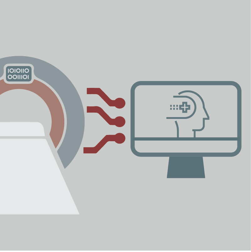
Medical image analysis holds immense value in modern healthcare, offering potential to revolutionize diagnostics, treatment planning, and patient care. By leveraging advanced technologies such as machine learning and computer vision, medical image analysis allows for precise and efficient interpretation of complex visual data from various imaging modalities like X-rays, Magnetic resonance imaging (MRI), and Computed Tomography scan (CT) scans.
Dive into the dynamic realm of medical image analysis with our introductory 2-day course, tailored to match the rapid pace of both the field and its foundational technology—computing. Structured to be practical and contemporary, the course takes a ‘hands-on’ approach, guiding participants from introductory imaging concepts through the development of compact machine learning pipelines for inference. By the workshop’s conclusion, attendees will not only have acquired a nuanced understanding of medical imaging intricacies but will also possess the requisite tools for autonomous image analysis. Furthermore, participants will be introduced to key publicly-available resources, ensuring they depart with a valuable skill set poised to contribute to advancements in this crucial field.
Participants will gain practical experience and skills to be able to:
- Understand key differences in medical imaging modalities,
- Learn basic image processing techniques,
- Process raw clinical images into analysis ready formats,
- Locate and download publicly available imaging data sets,
- Extract imaging features and train machine learning models for clinical prediction,
- Familiarize yourself with auto-segmentation tools and build deep learning models for medical image segmentation.
Graduates, postgraduates, staff bioinformaticians, PIs, clinicians, and anyone working with or about to embark on analysis and/or data processing of medical images.
Participants should be comfortable writing and reading basic Python. Scripting will be primarily in Google Colab notebook; it is recommended that participants have a Google account and a computer that can run the latest Google Chrome.
This workshop requires participants to complete pre-workshop tasks and readings.
Module 1: Introduction to medical imaging (Caryn Geady)
- Basic introduction to medical imaging and its applications
- Compare and contrast different medical imaging modalities
- Key image processing techniques
Lab Practical: Image Processing Fundamentals (Caryn Geady)
- Loading and querying images;
- Image processing including but not limited to:
- Histogram processing
- Spatial filtering
- Geometric transformations
Module 2: Hands-on with medical imaging (Caryn Geady, Sejin Kim)
- World coordinate vs image coordinate
- What is a DICOM? The glue that holds the coordinates together!
- Processing DICOM to analysis-ready images
- 2D vs 3D vs 4D images
Lab Practical: DICOM Wrestling (Caryn Geady, Ruiyan Ni, Sejin Kim)
- How to download a public dataset from The Cancer Imaging Archive (TCIA)
- Loading images with SimpleITK
- Manipulating imaging arrays in NumPy/Torch
- Data augmentation on 2D/3D images using TorchIO
Module 3: Machine learning applications I – Prediction (Sejin Kim)
- Imaging “biomarkers”
- Garbage-in-garbage-out! Gold-in-gold-out?: How to QC imaging data
- Radiomic feature extractions
- Building predictive models using deep learning
Lab Practical: Predictive Modeling (Sejin Kim)
- Cleaning your imaging data before analysis
- Using PyRadiomics to extract radiomic features
- Building basic machine learning models using Scikit-learn
- Building advanced deep learning models using PyTorch
- How to debug your models: finding model biases and pitfalls
Module 4: Machine learning applications II – Segmentation (Ruiyan Ni)
- Object detection vs instance segmentation vs semantic segmentation
- Exploring tools used to annotate images for label generation
- Learning traditional methods: Atlas-based, thresholding-based, and watershed-based methods
- Segmentation with deep learning
Lab Practical: Deep Learning Segmentation (Ruiyan Ni)
- Using traditional, non-machine learning method to segment an image
- Experiencing interactive segmentation using deep learning: ScribblePrompt method based on Segment Anything Model
- Building a basic U-Net model for 2D image segmentation by yourself
Duration: 2 days
Start: Sep 19, 2024
End: Sep 20, 2024
Status: Cancelled
Workshop Ended

Canadian Bioinformatics Workshops promotes open access. Past workshop content is available under a Creative Commons License.
Posted on:
 Download the poster announcing this workshop


